The first time you see a brain scan up close, it feels almost intrusive. All those folds and shadows, bright patches lighting up like a city at night. But here’s the thing most people miss: not all brain images are trying to answer the same question. Some ask, What does the brain look like? Others want to know, What is it doing right now? That split—between structural and functional brain imaging—is at the heart of modern neuroscience, medicine, and even psychology.
What Structural Brain Imaging Really Shows
Structural brain imaging is, at its core, anatomy. Think of it as a high-resolution map of the brain’s physical layout—its size, shape, and internal architecture. These scans don’t care what you’re thinking about or whether you’re stressed or calm. They’re frozen snapshots, like aerial photos taken from a satellite.
The most common tools here are MRI (Magnetic Resonance Imaging) and CT (Computed Tomography) scans. MRI, in particular, has become the gold standard because it can distinguish between gray matter, white matter, and cerebrospinal fluid with remarkable clarity.
Doctors rely on structural imaging to detect tumors, brain injuries, strokes, and degenerative diseases. A neurologist looking for multiple sclerosis lesions or early signs of Alzheimer’s isn’t asking about brain activity—they’re looking for physical changes, thinning tissue, or abnormal growths. The National Institute of Neurological Disorders and Stroke explains how MRI helps identify these structural abnormalities in clinical settings (https://www.ninds.nih.gov).
How Structural Imaging Works Under the Hood
MRI scanners don’t actually “see” the brain in the way a camera does. They use powerful magnetic fields and radio waves to measure how hydrogen atoms behave inside the body. Since different tissues contain different amounts of water and fat, they respond differently, allowing the scanner to construct a detailed image layer by layer.
CT scans, by contrast, rely on X-rays and are faster but less detailed for soft tissue. They’re often used in emergencies—head trauma, suspected bleeding—where speed matters more than nuance. According to the U.S. Food and Drug Administration, CT imaging remains critical for acute neurological diagnosis despite advances in MRI (https://www.fda.gov).
Functional Brain Imaging: Watching the Brain in Action
If structural imaging is a photograph, functional imaging is a movie. It captures changes over time, tracking which areas of the brain are active during specific tasks, emotions, or states of rest.
Functional MRI (fMRI) is the most widely known technique here. Instead of measuring anatomy, it tracks blood oxygen levels. When neurons fire, they consume oxygen. Blood flow increases to meet that demand, and fMRI detects these subtle shifts. That’s how scientists can say, with reasonable confidence, that a certain brain region “lights up” when you read, remember a face, or feel fear.
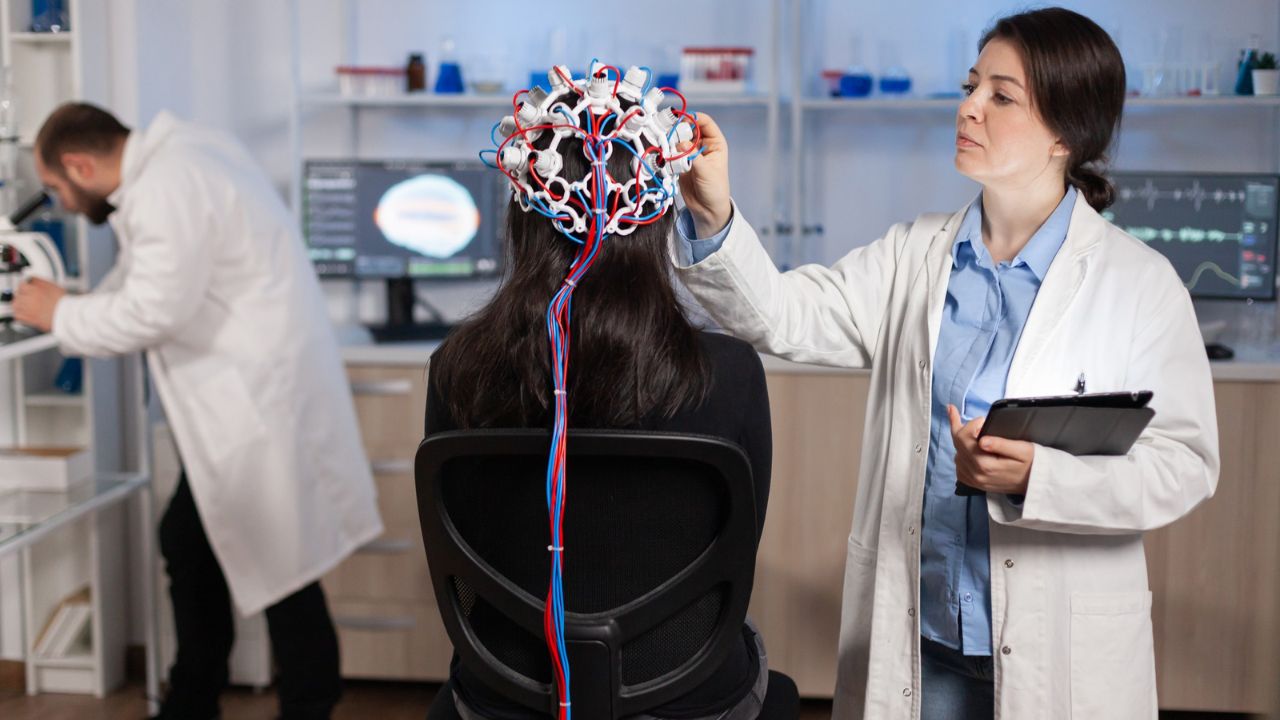
Other functional tools include PET (Positron Emission Tomography) scans and EEG (electroencephalography). PET scans use radioactive tracers to observe metabolic processes, while EEG measures electrical activity directly via electrodes on the scalp. Each has strengths and trade-offs, as outlined by the National Institutes of Health (https://www.nih.gov).
Structural vs Functional: A Side-by-Side Look
| Feature | Structural Imaging | Functional Imaging |
|---|---|---|
| Primary Question | What does the brain look like? | What is the brain doing? |
| Common Tools | MRI, CT | fMRI, PET, EEG |
| Time Sensitivity | Static snapshot | Dynamic, time-based |
| Clinical Use | Tumors, injuries, degeneration | Brain activity, cognition, behavior |
| Resolution | High spatial detail | Lower spatial, higher temporal detail |
This distinction matters more than it sounds. A perfectly normal-looking brain can function abnormally, and an abnormal-looking brain can sometimes function just fine. One doesn’t replace the other.
Why Researchers Often Use Both Together
Modern neuroscience rarely relies on a single imaging method. A researcher studying epilepsy might use structural MRI to locate damaged tissue, then fMRI or EEG to observe seizure activity. In psychiatric research, structural scans can reveal volume differences in certain regions, while functional scans show altered communication patterns between networks.
This combined approach is especially important in brain disorders where symptoms don’t map neatly onto anatomy. Depression, anxiety, and schizophrenia often show subtle structural changes, but more pronounced functional disruptions. The National Institute of Mental Health has emphasized the importance of multi-modal imaging in understanding these conditions (https://www.nimh.nih.gov).
Limits, Misinterpretations, and Media Hype
Here’s where things get tricky. Functional brain images, especially those colorful fMRI maps, are often misunderstood. Those glowing reds and yellows don’t mean a brain region is “working harder” in a simple sense. They reflect statistical differences in blood flow, processed through layers of analysis and assumptions.
Structural imaging has its own limitations. A visible abnormality doesn’t always explain a patient’s symptoms, and “normal” scans don’t rule out serious dysfunction. The brain is messy, adaptive, and deeply individual. That’s something both clinicians and journalists have had to relearn over the years.
The Future of Brain Imaging Science
New techniques are blurring the line between structure and function. Diffusion tensor imaging (DTI), for example, maps white matter pathways, revealing how different regions are physically connected. Resting-state fMRI studies how brain networks communicate even when you’re doing nothing at all.
Artificial intelligence is also stepping in, helping researchers detect patterns too subtle for the human eye. According to recent updates from the National Library of Medicine, machine learning models are already improving diagnostic accuracy in neuroimaging research (https://www.nlm.nih.gov).
Why This Distinction Matters Beyond the Lab
Understanding the difference between structural and functional imaging isn’t just academic. It shapes how diseases are diagnosed, how treatments are evaluated, and how we interpret claims about the brain in popular media. When someone says, “Scientists found the brain region responsible for happiness,” the first question should be: Using what kind of imaging?
Structural scans tell us where the brain has been altered. Functional scans tell us how it behaves in the moment. Together, they offer a fuller—though still incomplete—picture of the most complex organ we have.
In the end, brain imaging doesn’t give us definitive answers. It gives us better questions. And in neuroscience, that’s often where the real progress begins.
FAQs:
Is functional brain imaging more accurate than structural imaging?
They serve different purposes. Functional imaging isn’t “more accurate”; it answers different questions about brain activity rather than anatomy.
Can a structural MRI diagnose mental illness?
Not on its own. Structural changes may support a diagnosis, but mental illnesses are primarily diagnosed through clinical evaluation.
Why are fMRI images so colorful?
The colors represent statistical differences in blood oxygen levels, not direct measurements of thoughts or emotions.