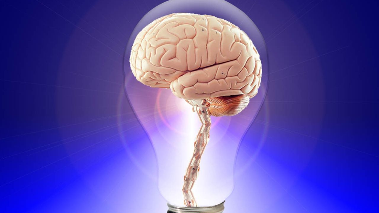
Early detection is one of the most powerful tools in modern medicine, especially when it comes to neurological disorders. Conditions such as Alzheimer’s disease, Parkinson’s disease, multiple sclerosis, epilepsy, stroke, and brain tumors often begin developing years before clear symptoms appear. Brain imaging research plays a crucial role in identifying these changes early—sometimes even before patients notice anything is wrong.
By allowing doctors and researchers to see inside the brain noninvasively, imaging technologies have transformed how neurological diseases are diagnosed, monitored, and treated.
Why Early Detection Matters
Neurological disorders are often progressive, meaning they worsen over time. Once significant brain damage occurs, it is usually irreversible. Early detection allows:
- Earlier medical intervention
- Slower disease progression
- Better symptom management
- Improved quality of life
- More accurate treatment planning
In diseases like Alzheimer’s, early diagnosis can add years of independent living. For stroke patients, rapid brain imaging can mean the difference between recovery and permanent disability.
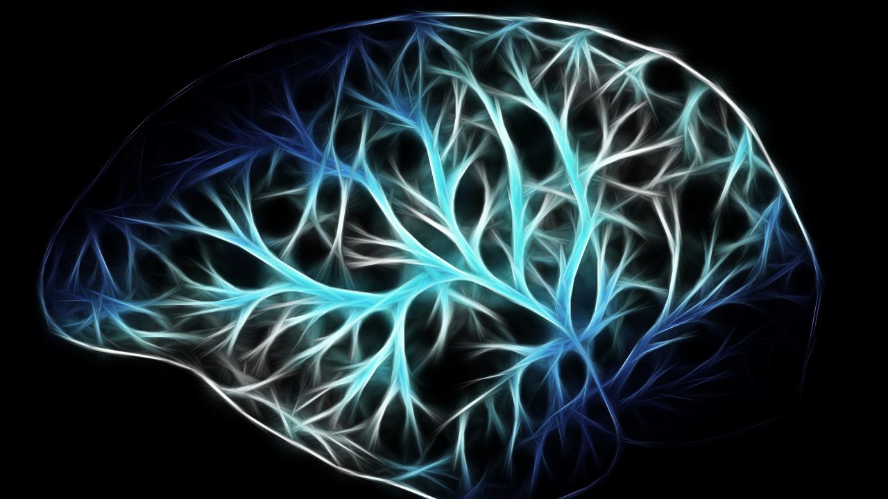
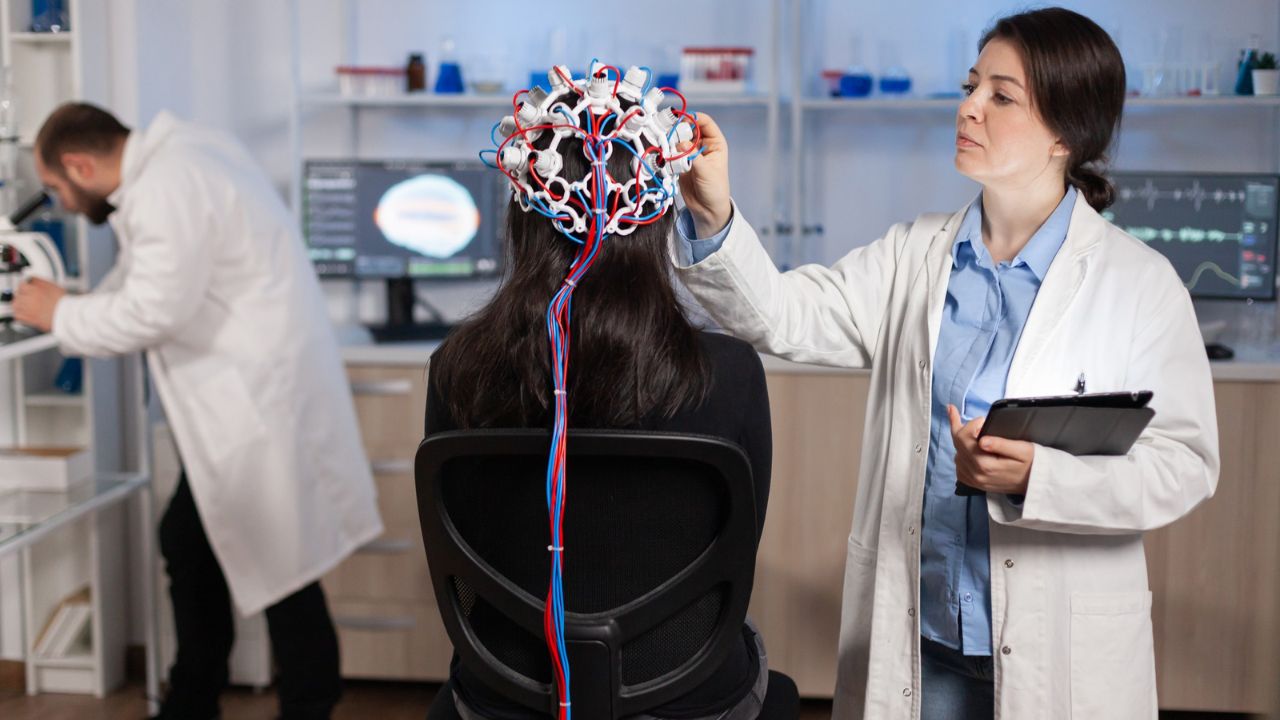
Key Brain Imaging Technologies Used in Early Detection
Modern neuroscience relies on several imaging tools, each providing different types of information about brain structure, function, and chemistry.
Magnetic Resonance Imaging (MRI)
MRI is one of the most widely used tools for detecting structural brain abnormalities. It can identify:
- Brain atrophy (shrinking), common in Alzheimer’s disease
- Lesions associated with multiple sclerosis
- Tumors, bleeding, or swelling
- Early signs of stroke
High-resolution MRI scans allow clinicians to detect subtle brain changes years before symptoms become severe.
Functional MRI (fMRI)
fMRI measures brain activity by detecting changes in blood flow. It is particularly useful for:
- Identifying early functional changes in Alzheimer’s and dementia
- Detecting abnormal brain network activity in epilepsy
- Studying cognitive decline and memory disorders
Even when brain structure looks normal, fMRI can reveal altered brain function—often an early warning sign of disease.
Positron Emission Tomography (PET)
PET scans are essential for detecting biochemical changes in the brain. They are commonly used to:
- Identify amyloid plaques and tau proteins linked to Alzheimer’s disease
- Detect reduced dopamine activity in Parkinson’s disease
- Diagnose brain cancers and assess tumor spread
PET imaging often detects disease before structural damage is visible on MRI or CT scans.
Computed Tomography (CT)
CT scans are fast and widely available, making them critical in emergency settings. They are often used to:
- Quickly diagnose strokes or brain hemorrhages
- Detect traumatic brain injuries
- Identify tumors or swelling
While CT is less detailed than MRI, its speed makes it vital for early intervention in acute neurological conditions.
Disorders Commonly Detected Early Through Brain Imaging
Brain imaging has significantly improved early diagnosis across many neurological conditions:
- Alzheimer’s disease: Brain shrinkage, amyloid buildup, reduced metabolic activity
- Parkinson’s disease: Dopamine system dysfunction via PET scans
- Multiple sclerosis: Early white-matter lesions visible on MRI
- Epilepsy: Abnormal brain activity patterns and focal lesions
- Stroke: Blocked or ruptured blood vessels detected immediately
- Brain tumors: Early mass detection before neurological symptoms appear
Benefits for Treatment and Research
Early detection through brain imaging doesn’t just help patients—it advances science. Imaging allows researchers to:
- Track disease progression over time
- Test the effectiveness of new drugs
- Identify high-risk individuals for preventive care
- Develop personalized treatment strategies
This has accelerated clinical trials and improved outcomes by matching therapies to individual brain patterns.
Limitations and Challenges
Despite its power, brain imaging has limitations:
- High costs and limited access in some regions
- Incidental findings that may cause anxiety
- Need for expert interpretation
- Not all brain changes guarantee disease development
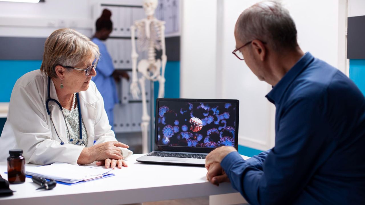
For this reason, imaging is often combined with clinical exams, genetic testing, and cognitive assessments.
The Future of Early Detection
The future of brain imaging lies in artificial intelligence and machine learning. AI algorithms can analyze scans faster and detect patterns invisible to the human eye. Combined with wearable devices and genetic data, brain imaging may soon predict neurological disorders years before symptoms appear.
FAQs:
What is brain imaging?
Brain imaging refers to medical techniques that create visual representations of the brain’s structure, function, or chemistry to help diagnose and study neurological conditions.
Why is brain imaging important for early detection?
Brain imaging can reveal changes in the brain before symptoms appear, allowing earlier diagnosis, faster treatment, and better long-term outcomes.
Which brain imaging test is best for early diagnosis?
There is no single “best” test. MRI is widely used for structural changes, fMRI for brain activity, PET for chemical changes, and CT scans for emergencies like strokes or head injuries.