Medical imaging has transformed modern healthcare by allowing doctors and researchers to see inside the human body without surgery. Among the most important imaging tools used today are MRI, fMRI, PET, and CT scans. While these technologies are often mentioned together, each serves a distinct purpose and provides different types of information about the body—especially the brain.
This guide explains how each scan works, what it is used for, and how they differ, helping patients and readers better understand these critical diagnostic tools.
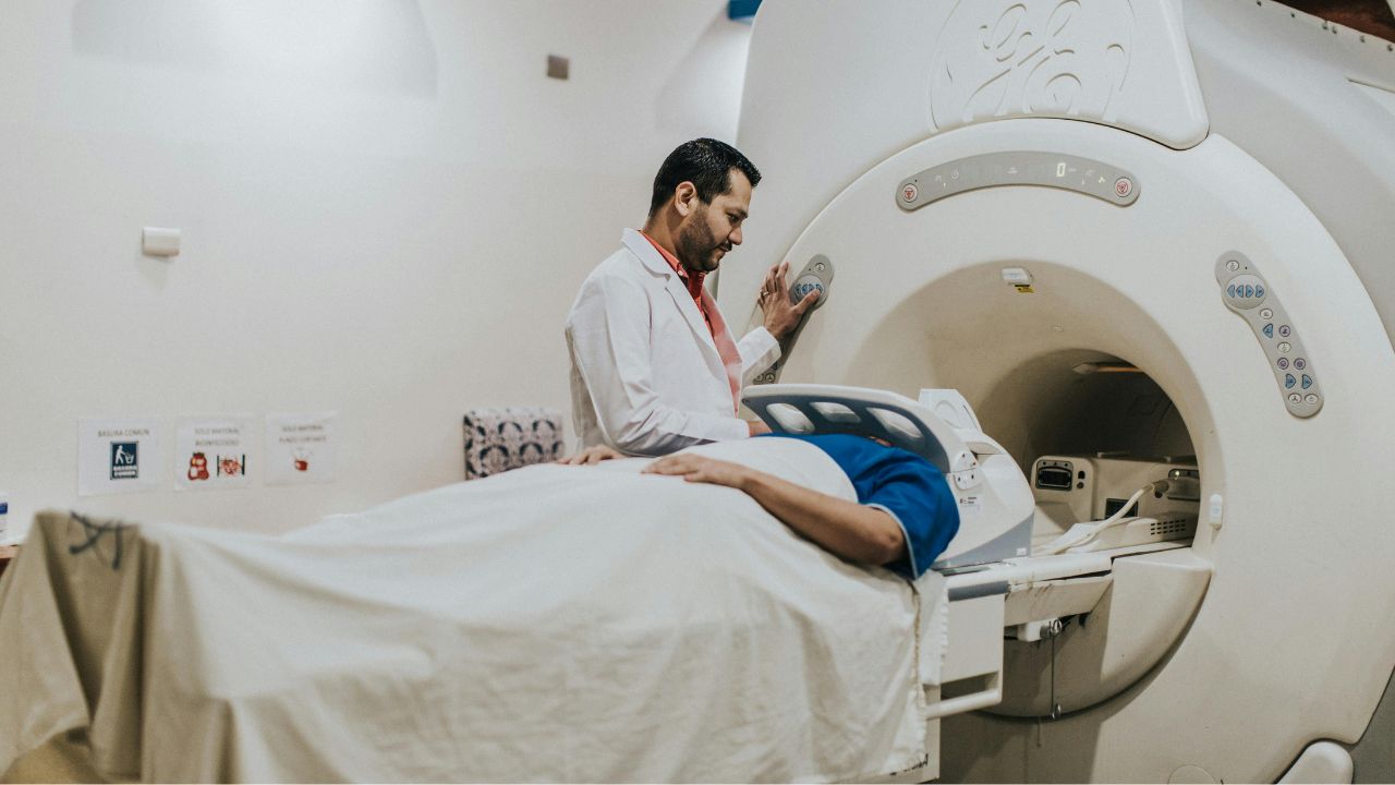
What Is MRI (Magnetic Resonance Imaging)?
MRI, or Magnetic Resonance Imaging, uses strong magnetic fields and radio waves to create highly detailed images of organs and tissues.
How MRI Works
MRI scanners align hydrogen atoms in the body using a powerful magnet. Radiofrequency pulses then disturb this alignment, and as atoms return to their original state, they emit signals that are converted into images by a computer.
What MRI Is Used For
- Brain and spinal cord disorders
- Tumors and cysts
- Joint and muscle injuries
- Heart and blood vessel abnormalities
- Soft tissue imaging
Key Advantages
- No ionizing radiation
- Extremely detailed images of soft tissue
- Safe for repeated use (with some metal-related exceptions)
Limitations
- Expensive
- Loud and time-consuming
- Not suitable for patients with certain implants or severe claustrophobia
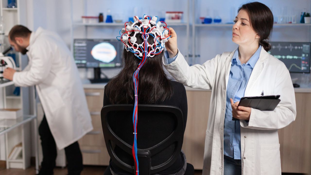
What Is fMRI (Functional Magnetic Resonance Imaging)?
fMRI is a specialized type of MRI that measures brain activity rather than just structure.
How fMRI Works
fMRI detects changes in blood oxygen levels. When a region of the brain becomes active, it consumes more oxygen, and blood flow to that area increases. These changes are captured and mapped in real time.
What fMRI Is Used For
- Studying brain function
- Mapping language, memory, and motor areas
- Pre-surgical brain planning
- Cognitive and behavioral neuroscience research
- Mental health research
Key Advantages
- Non-invasive
- No radiation
- Shows brain activity in real time
Limitations
- Sensitive to movement
- Does not directly measure neuron firing
- Mostly used in research and specialized clinical cases
What Is PET (Positron Emission Tomography)?
PET scans show how organs and tissues function at the cellular level using radioactive tracers.
How PET Works
A small amount of radioactive tracer is injected into the body. As the tracer breaks down, it emits positrons that collide with electrons, producing signals detected by the scanner.
What PET Is Used For
- Cancer detection and staging
- Brain disorders like Alzheimer’s and Parkinson’s
- Epilepsy evaluation
- Heart disease assessment
- Measuring metabolism and blood flow
Key Advantages
- Detects disease before structural changes appear
- Shows metabolic and chemical activity
- Highly sensitive for cancer and neurological conditions
Limitations
- Uses radiation
- Expensive
- Less detailed anatomical images (often combined with CT or MRI)
What Is CT (Computed Tomography) Scan?
CT scans use X-rays and computer processing to create cross-sectional images of the body.
How CT Works
An X-ray tube rotates around the body, capturing multiple images from different angles. These images are combined to form detailed slices of internal structures.
What CT Is Used For
- Head injuries and strokes
- Internal bleeding
- Bone fractures
- Lung and chest imaging
- Emergency trauma assessment
Key Advantages
- Fast and widely available
- Excellent for detecting bone injuries and bleeding
- Ideal for emergency situations
Limitations
- Uses ionizing radiation
- Less detailed soft-tissue images than MRI
- Not ideal for frequent repeat scans
Comparing MRI, fMRI, PET, and CT Scans
| Feature | MRI | fMRI | PET | CT |
|---|---|---|---|---|
| Radiation | No | No | Yes | Yes |
| Main Focus | Structure | Brain activity | Metabolism | Structure |
| Scan Time | Long | Long | Moderate | Short |
| Best For | Soft tissues | Brain function | Cancer & brain disorders | Trauma & emergencies |
| Cost | High | High | Very high | Moderate |
Which Scan Is Right for You?
The choice depends on medical need:
- MRI: Best for detailed structural imaging of soft tissues
- fMRI: Ideal for brain function and cognitive studies
- PET: Used when metabolic or chemical activity needs evaluation
- CT: Preferred for emergencies and quick diagnosis
Doctors often combine scans (such as PET-CT or MRI-fMRI) to get a complete picture.
FAQs:
What is the main difference between MRI and CT scans?
The main difference lies in how images are created. MRI uses magnetic fields and radio waves, while CT scans use X-rays. MRI is better for soft tissues like the brain and muscles, whereas CT scans are faster and often used in emergencies to detect fractures or internal bleeding.
Does an MRI or fMRI expose patients to radiation?
No. Neither MRI nor fMRI uses ionizing radiation. Both are considered safe for repeated use, as long as the patient does not have metal implants that are incompatible with MRI scanners.
What makes fMRI different from a regular MRI?
MRI shows the structure of the brain, while fMRI shows how the brain functions. fMRI tracks changes in blood oxygen levels to identify which brain regions are active during tasks like speaking, thinking, or moving.