It often starts quietly. A missed appointment. A familiar name stuck just out of reach. By the time Alzheimer’s disease announces itself clinically, the brain has already been changing for years—sometimes decades. That gap between invisible damage and visible symptoms is where brain imaging research has reshaped the entire dementia field, turning guesswork into measurable biology.
Today, brain scans aren’t just confirming dementia after the fact. They’re helping scientists track its earliest footprints, understand why it spreads the way it does, and test whether treatments are actually slowing the disease—or just buying time.
Seeing Alzheimer’s Before Memory Fails
For most of the 20th century, Alzheimer’s could only be definitively diagnosed after death, through autopsy. Imaging changed that narrative.
Structural MRI was the first major breakthrough. Researchers noticed consistent patterns of brain atrophy, particularly in the hippocampus and surrounding medial temporal lobe regions—areas essential for memory formation. These weren’t random shrinkages. They followed a recognizable progression.
Longitudinal MRI studies showed that people who later developed Alzheimer’s began losing volume years before symptoms emerged. According to the National Institute on Aging, these structural changes are now considered core biological markers of the disease (https://www.nia.nih.gov).
This shifted dementia research from reaction to prediction.
Amyloid and Tau: Imaging the Hallmarks of Disease
The real paradigm shift came with molecular imaging. PET scans, using specialized radioactive tracers, allowed researchers to visualize amyloid-beta plaques and tau tangles inside living brains—once thought impossible.
Amyloid PET imaging revealed a sobering truth: many cognitively normal older adults already carry significant amyloid buildup. This forced researchers to rethink timelines. Alzheimer’s wasn’t a sudden event. It was a long, slow biological process.
Tau PET imaging added even more precision. Unlike amyloid, tau accumulation correlates closely with symptom severity and cognitive decline. Studies supported by the National Institutes of Health show that tau spread aligns with the step-by-step worsening of dementia (https://www.nih.gov).
In research settings, amyloid tells you who is at risk. Tau tells you who is declining.
Structural MRI and Brain Atrophy Patterns
Structural imaging remains the backbone of dementia research. MRI doesn’t just show shrinkage; it reveals where degeneration starts and how it spreads.
Alzheimer’s follows a fairly predictable pattern, beginning in memory-related regions before advancing to language, spatial, and executive networks. Other dementias look different. Frontotemporal dementia targets frontal and temporal lobes. Vascular dementia shows damage linked to blood flow and microinfarcts.
These distinctions matter. Imaging helps researchers differentiate between dementia subtypes that often look similar clinically but require different management strategies. The National Institute of Neurological Disorders and Stroke emphasizes MRI’s role in distinguishing Alzheimer’s from other neurodegenerative diseases (https://www.ninds.nih.gov).
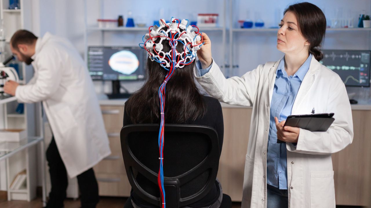
Functional Imaging and Failing Brain Networks
While structural MRI shows damage, functional imaging shows dysfunction. fMRI studies in Alzheimer’s research often focus on disrupted communication between brain regions rather than isolated failures.
One of the earliest functional changes appears in the default mode network—the same system involved in memory and self-referential thought. Even before significant atrophy, this network begins to fragment.
FDG-PET scans, which measure glucose metabolism, have been especially useful. Reduced metabolic activity in temporoparietal regions often precedes noticeable cognitive decline. According to the U.S. Food and Drug Administration, FDG-PET is an accepted supportive tool in dementia research and differential diagnosis (https://www.fda.gov).
In short, the brain may still look intact, but it’s already running on low power.
Mild Cognitive Impairment: The Imaging Battleground
Mild cognitive impairment (MCI) sits in the gray zone between normal aging and dementia. Not everyone with MCI progresses to Alzheimer’s—but many do.
Brain imaging has become central to sorting that uncertainty. Individuals with MCI who show hippocampal atrophy, amyloid positivity, and tau accumulation face a significantly higher risk of progression.
Large-scale studies like the Alzheimer’s Disease Neuroimaging Initiative (ADNI) have combined MRI, PET, genetics, and cognitive testing to refine predictive models. Data from ADNI, funded by the NIH, has shaped nearly every major Alzheimer’s imaging study of the past decade (https://adni.loni.usc.edu).
This is where imaging moves from descriptive to decisional.
Imaging’s Role in Drug Development
Few areas have benefited more from brain imaging than Alzheimer’s drug research—especially after decades of failed clinical trials.
Modern trials now require biological confirmation of disease using imaging biomarkers. Amyloid PET scans ensure participants actually have Alzheimer’s pathology before enrollment. MRI monitors brain volume changes to detect potential side effects like swelling or bleeding.
When controversial anti-amyloid drugs entered the market, imaging was central to both approval and debate. Regulators relied heavily on PET evidence showing plaque reduction, even as questions remained about clinical benefit. The FDA has publicly detailed how imaging biomarkers factor into Alzheimer’s drug evaluation (https://www.fda.gov).
Without imaging, these trials would be flying blind.
Beyond Alzheimer’s: Other Dementias in Focus
Brain imaging research has expanded well beyond Alzheimer’s alone. Lewy body dementia shows characteristic patterns on dopamine transporter scans. Vascular dementia reveals white matter damage tied to chronic blood vessel disease. Normal pressure hydrocephalus presents with enlarged ventricles—often reversible if caught early.
These distinctions matter because treatments differ dramatically. Imaging doesn’t just label disease; it opens doors to interventions that might otherwise be missed.
Artificial Intelligence and the Next Frontier
The newest wave of dementia imaging research leans heavily on artificial intelligence. Machine learning algorithms can detect subtle patterns of atrophy and connectivity loss long before human readers can.
According to recent reviews from the National Library of Medicine, AI-driven analysis of brain scans is improving early detection accuracy and helping identify distinct Alzheimer’s subtypes (https://www.nlm.nih.gov).
This raises the possibility—still controversial—that one day a routine brain scan could flag dementia risk years in advance. The ethical questions are as big as the technical ones.
Limits, Risks, and Hard Truths
Despite its power, brain imaging isn’t destiny. Amyloid-positive individuals may never develop dementia. Some patients decline rapidly with relatively modest imaging changes.
Cost, access, and radiation exposure (in PET scans) remain barriers. And no scan can yet predict exactly when symptoms will appear or how fast decline will occur.
The Alzheimer’s Association has repeatedly cautioned against overinterpreting imaging findings outside research settings (https://www.alz.org). Context still matters. So does the human story behind the scan.
Why Imaging Has Changed the Dementia Narrative
Brain imaging hasn’t cured Alzheimer’s. But it has fundamentally altered how the disease is understood. Dementia is no longer an abrupt clinical event—it’s a long biological process unfolding silently over time.
That realization has redirected research dollars, reshaped clinical trials, and given patients and families something rare in this field: earlier answers.
In Alzheimer’s research, the brain scan isn’t just a picture. It’s a timeline. And for a disease defined by memory loss, that may be its most valuable contribution yet.
FAQs:
Can brain imaging diagnose Alzheimer’s definitively?
Imaging can strongly support a diagnosis but is usually combined with clinical evaluation and cognitive testing.
What scan is best for detecting early Alzheimer’s?
Amyloid and tau PET scans are most specific, while MRI detects early structural changes.
Are brain scans used routinely for dementia diagnosis?
Not always. Cost and availability limit routine use, especially for PET imaging.