A traumatic brain injury rarely announces its full damage on day one. The CT scan might look “clean.” The patient might even walk out of the emergency room. Weeks or months later, though, headaches linger, memory slips, emotions tilt off balance, and something clearly isn’t right. That disconnect—between visible injury and lived experience—is exactly why brain imaging has become such a central force in traumatic brain injury (TBI) research.
In TBI, imaging isn’t just about spotting damage. It’s about uncovering what standard scans miss, tracking invisible injuries, and understanding how the brain struggles, adapts, and sometimes heals after trauma.
Why TBI Is So Hard to See
Unlike stroke or tumors, many TBIs don’t leave behind dramatic structural scars. Especially in mild TBI or concussion, the injury is often diffuse, microscopic, and network-based. Neurons stretch. Axons shear. Connections misfire. None of that shows up cleanly on routine scans.
For years, this gap fueled skepticism. Patients reported symptoms. Imaging said “normal.” Research eventually caught up to what patients already knew: absence of evidence isn’t evidence of absence.
The National Institute of Neurological Disorders and Stroke acknowledges that conventional imaging often underestimates TBI severity, particularly in mild cases (https://www.ninds.nih.gov). That realization reshaped the field.
CT Scans: Life-Saving but Limited
In the acute phase, computed tomography (CT) is king. It’s fast, accessible, and excellent at detecting bleeding, skull fractures, and life-threatening swelling.
In emergency care, CT answers urgent questions: Is there hemorrhage? Is surgery needed now? Is intracranial pressure rising?
But for research—and long-term understanding—CT hits a wall. Many patients with persistent post-concussive symptoms show normal CT scans. That doesn’t mean their brains are uninjured. It means the injury lives below CT’s resolution.
CT saves lives. It doesn’t tell the whole story.
Structural MRI: Finding Subtle Damage
MRI pushed TBI research forward by revealing injuries CT couldn’t. Contusions, microbleeds, and subtle tissue loss became visible with higher resolution imaging.
Advanced MRI sequences like susceptibility-weighted imaging (SWI) detect tiny hemorrhages associated with diffuse axonal injury (DAI)—a hallmark of moderate to severe TBI. These microbleeds often correlate with worse cognitive outcomes.
Longitudinal MRI studies track brain volume loss over time, showing that TBI can trigger progressive neurodegeneration long after the initial impact. According to the National Institutes of Health, this delayed atrophy helps explain why symptoms may worsen months or years after injury (https://www.nih.gov).
In TBI research, MRI revealed that damage isn’t always immediate—and recovery isn’t always linear.
Diffuse Axonal Injury and DTI
If there’s one imaging tool that transformed TBI research, it’s diffusion tensor imaging (DTI). DTI maps white matter tracts—the brain’s communication cables—by tracking water movement along axons.
Traumatic forces stretch and shear these fibers, disrupting communication even when neurons remain alive. DTI can detect reduced white matter integrity that standard MRI misses entirely.
These findings validated what many patients experienced: slowed thinking, poor attention, emotional volatility. The wiring was compromised, not destroyed.
Studies summarized by the National Library of Medicine show strong links between DTI abnormalities and cognitive deficits after TBI (https://www.nlm.nih.gov). This moved the field from “invisible injury” to measurable biology.
Functional MRI and Network Disruption
Structural damage is only part of the picture. Functional MRI (fMRI) reveals how brain networks behave after injury—and how they compensate.
In TBI, fMRI studies often show abnormal activation patterns. Tasks that require minimal effort in healthy brains may demand widespread recruitment after injury. The brain works harder to achieve the same outcome.
Resting-state fMRI has been particularly revealing. Even when patients are lying still, their brain networks show altered connectivity, especially in attention, executive control, and default mode networks.
These disruptions persist even after symptoms improve, suggesting that recovery often involves compensation rather than full restoration. The National Institute of Mental Health has supported network-based imaging models to better understand these changes (https://www.nimh.nih.gov).
Mild TBI and Concussion: Imaging the Gray Zone
Mild TBI accounts for the vast majority of brain injuries—and the most controversy. Athletes, military personnel, and accident victims often show lingering symptoms with “normal” scans.
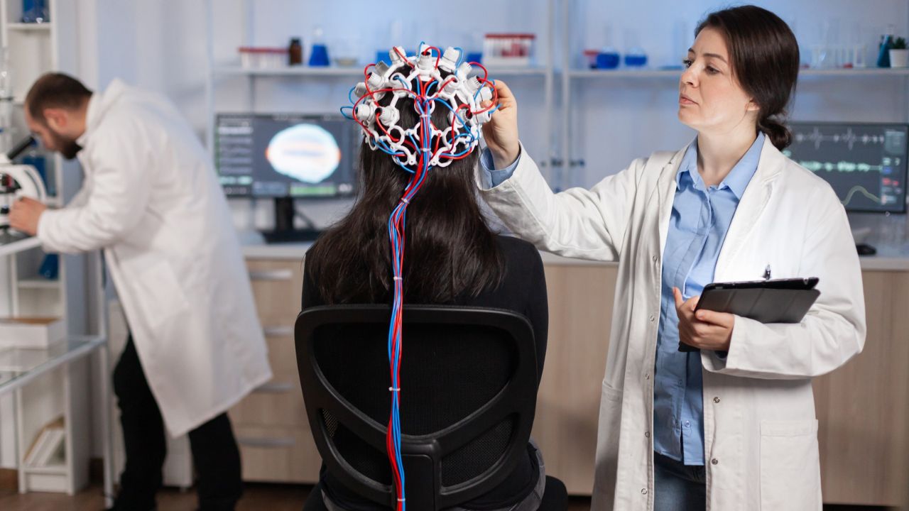
Advanced imaging changed that conversation. DTI, fMRI, and EEG studies consistently reveal subtle but reproducible abnormalities after concussion, even when standard MRI looks normal.
This doesn’t mean every concussion leaves permanent damage. But it does mean that repeated injuries, insufficient recovery time, and individual vulnerability matter.
The Centers for Disease Control and Prevention has emphasized that imaging findings support stricter concussion management and return-to-play guidelines (https://www.cdc.gov).
In research terms, imaging helped legitimize concussion as a real brain injury—not just a temporary inconvenience.
Imaging Recovery and Brain Plasticity
One of the most hopeful roles of brain imaging in TBI research is tracking recovery.
Longitudinal studies show that brain networks can reorganize after injury. Functional imaging reveals shifts in activation patterns as patients regain skills. White matter integrity may partially recover, especially with targeted rehabilitation.
Imaging has helped identify biomarkers of good recovery—preserved tracts, adaptive network reorganization, and early normalization of connectivity. These markers guide both prognosis and therapy development.
NIH-funded studies demonstrate that rehabilitation changes brain activity, not just behavior (https://www.nih.gov). In TBI, therapy doesn’t just teach coping strategies—it reshapes the brain.
TBI, Mental Health, and Imaging Links
Depression, anxiety, irritability, and impulse control problems are common after TBI. Imaging studies reveal why.
Damage to frontal-limbic circuits—often subtle—disrupts emotional regulation. Functional imaging shows altered activity in networks involved in mood, reward, and stress response.
These findings helped dismantle the false divide between “psychological” and “organic” symptoms. After TBI, mental health changes are neurological changes.
The Department of Veterans Affairs has relied heavily on imaging research to understand TBI-related mental health outcomes in service members (https://www.ptsd.va.gov).
Chronic Traumatic Encephalopathy and Long-Term Imaging
CTE remains a diagnosis made after death, but imaging research is closing the gap. PET imaging targeting tau protein is being studied as a potential marker of chronic injury in athletes and military populations.
Structural MRI studies show progressive atrophy patterns in individuals with repeated head trauma. While no scan can yet diagnose CTE definitively, imaging is shaping risk models and prevention strategies.
This is one area where caution is essential. The science is evolving, and overinterpretation carries real ethical risks.
Artificial Intelligence in TBI Imaging
AI is rapidly reshaping TBI imaging research. Machine learning models analyze complex patterns across MRI, DTI, and fMRI data to predict outcomes, classify injury severity, and identify patients at risk for prolonged recovery.
These tools are particularly valuable in mild TBI, where differences are subtle and distributed. According to recent NIH-supported reviews, AI-assisted analysis improves sensitivity without sacrificing specificity (https://www.nlm.nih.gov).
The promise is personalization: matching rehabilitation intensity, timing, and strategies to an individual brain’s injury pattern.
Limits and Cautions
Despite its advances, brain imaging cannot diagnose TBI in isolation. Findings overlap with normal variation, stress effects, and pre-existing conditions.
Access remains uneven. Advanced imaging is expensive and not universally available. Interpretation requires expertise. And scans can’t measure pain, fatigue, or lived experience.
The American College of Radiology emphasizes that imaging should support—not override—clinical assessment in TBI care (https://www.acr.org).
Why Imaging Changed TBI Research
Brain imaging didn’t make TBI simpler. It made it more honest.
It showed that brain injury isn’t always obvious, that recovery isn’t binary, and that symptoms don’t require visible lesions to be real. It validated patients, refined diagnosis, and reshaped rehabilitation science.
In TBI research, imaging doesn’t just reveal damage. It reveals vulnerability, adaptation, and resilience—all unfolding in a brain trying to find its footing after impact.
That insight has changed everything.
FAQs:
Can brain imaging diagnose a concussion?
Not definitively. Imaging can show supportive findings but diagnosis remains clinical.
Why do many TBI scans look normal?
Because damage is often microscopic or network-based, beyond standard scan resolution.
What imaging is most useful for TBI research?
MRI, DTI, and fMRI are most informative beyond acute CT.