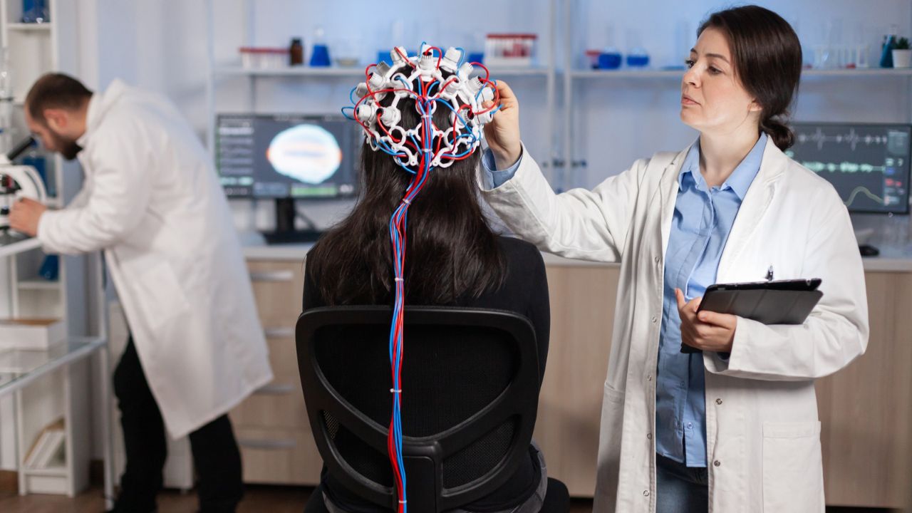
A decade ago, brain imaging analysis was slow, manual, and deeply human. Radiologists scrolled slice by slice. Researchers hand-labeled regions. Subtle patterns lived or died by the sharpness of the human eye. Today, that workflow feels almost antique. Artificial intelligence and machine learning have moved into the scanner room—and they’re not just speeding things up. They’re changing what scientists can see in the first place.
In brain imaging, AI isn’t replacing experts. It’s reshaping the limits of analysis, pushing neuroresearch from description toward prediction.
Why Brain Imaging Was Ripe for AI
Neuroimaging produces overwhelming amounts of data. A single MRI scan can contain millions of data points. Large research studies generate tens of thousands of scans across time, tasks, and populations.
Humans are good at recognizing known patterns. They’re not great at discovering hidden ones across massive, high-dimensional datasets. That gap is exactly where machine learning thrives.
According to the National Institutes of Health, advances in computational power and data availability have made AI-based neuroimaging analysis a priority area for federally funded research (https://www.nih.gov). The bottleneck was never data. It was interpretation.
From Handcrafted Features to Learned Patterns
Traditional brain imaging analysis relied on predefined features. Researchers decided what mattered—volume of a region, thickness of a cortex, average activation in a voxel—and tested hypotheses accordingly.
Machine learning flipped that model.
Instead of telling the algorithm what to look for, researchers train models on labeled data and let the system learn relevant patterns on its own. Deep learning models, especially convolutional neural networks (CNNs), can analyze raw imaging data with minimal preprocessing.
This matters because the brain doesn’t always change in neat, anatomically obvious ways. AI can detect distributed, subtle shifts across networks that escape conventional statistical methods.
The National Library of Medicine has documented how deep learning models outperform traditional approaches in tasks like lesion detection and disease classification (https://www.nlm.nih.gov).
Faster, More Accurate Image Processing
One of AI’s earliest wins in neuroimaging was automation.
Tasks that once took hours—or days—can now be completed in minutes. Brain segmentation, skull stripping, tissue classification, motion correction: these are foundational steps in imaging analysis, and they’re now largely AI-driven.
This speed isn’t just convenient. It reduces human variability and error. Two experts may draw slightly different boundaries around a brain structure. An algorithm, once validated, does it the same way every time.
The FDA has cleared multiple AI-based imaging tools that assist with image processing and abnormality detection, signaling growing regulatory confidence in these systems (https://www.fda.gov).
Detecting Disease Earlier Than Humans Can
Perhaps the most transformative impact of AI in brain imaging is early detection.
Machine learning models trained on thousands of scans can identify patterns associated with Alzheimer’s disease, Parkinson’s disease, multiple sclerosis, and stroke—sometimes years before symptoms appear.
In Alzheimer’s research, AI models analyzing MRI and PET scans have identified atrophy and metabolic patterns predictive of future cognitive decline. These signals are often too subtle or diffuse for visual inspection.
NIH-supported studies show that AI-assisted imaging analysis improves sensitivity for early neurodegenerative changes while maintaining clinical specificity (https://www.nih.gov).
That doesn’t mean diagnosis is automatic. It means the window for intervention is widening.
Functional Imaging and Brain Networks
Functional MRI generates especially complex data. It’s noisy, time-dependent, and network-based. This makes it a natural fit for machine learning.
AI models excel at identifying connectivity patterns across the brain—how regions synchronize, decouple, or reorganize during tasks or rest. These patterns are now central to research in mental health, autism, and recovery after brain injury.
Instead of asking whether one region is “overactive,” researchers can examine entire networks and how they shift under different conditions. Machine learning helps classify these network states and relate them to behavior or symptoms.
The National Institute of Mental Health has highlighted AI-based functional imaging analysis as key to advancing circuit-level models of psychiatric disorders (https://www.nimh.nih.gov).
Predicting Outcomes, Not Just Labeling Images
Traditional imaging answers descriptive questions: What does this scan show right now?
AI pushes toward predictive questions: What will happen next?
Machine learning models are being trained to predict stroke recovery, seizure recurrence, cognitive decline, and treatment response based on baseline imaging. These predictions are probabilistic, not deterministic—but they’re often better than chance and improving rapidly.
In stroke research, AI models combining imaging and clinical data now estimate tissue survival and functional recovery more accurately than time-based rules alone. The National Institute of Neurological Disorders and Stroke has supported this shift toward outcome-oriented imaging research (https://www.ninds.nih.gov).
Prediction is where imaging becomes decision-support rather than documentation.
Big Data, Shared Brains, and Open Science
AI thrives on scale, and neuroimaging has embraced data sharing. Large consortia pool brain scans from thousands of participants across countries and scanners.
Projects like the Alzheimer’s Disease Neuroimaging Initiative and UK Biobank created the kind of datasets machine learning needs to generalize beyond a single lab.
But scale brings challenges. Imaging protocols vary. Populations are uneven. Bias creeps in quietly.
The NIH has emphasized the need for diverse, representative imaging datasets to prevent AI models from learning narrow or misleading “norms” (https://www.nih.gov).
Bias, Black Boxes, and Trust
AI’s power comes with discomfort. Many machine learning models—especially deep neural networks—are difficult to interpret. They can make accurate predictions without explaining why.
In medicine, that’s a problem.
Clinicians and patients want transparency, not just performance. A model that flags high dementia risk without explanation raises ethical and practical concerns.
Bias is another issue. If training data underrepresents certain age groups, ethnicities, or neurological profiles, predictions may be less accurate—or unfair.
The FDA and NIH are both developing guidelines for explainability, validation, and post-deployment monitoring of AI-based medical imaging tools (https://www.fda.gov).
Accuracy alone is no longer enough.
AI as a Collaborator, Not a Replacement
Despite headlines, AI is not replacing radiologists or neuroscientists. It’s changing their role.
Experts increasingly act as supervisors, validators, and interpreters of algorithmic output. They decide when a model’s prediction makes sense—and when it doesn’t.
In research, AI generates hypotheses humans might not think to ask. In clinics, it flags cases that need urgent attention. The final judgment still rests with people.
That human-in-the-loop model is now considered best practice across neuroimaging research.
The Next Frontier: Multimodal Intelligence
The future of AI in brain imaging isn’t just better image analysis. It’s integration.
Researchers are combining imaging with genetics, blood biomarkers, wearable data, and electronic health records. Machine learning models can synthesize these signals into unified predictions.
This multimodal approach reflects reality. Brain disorders don’t live in scans alone. They live in biology, behavior, and environment.
The National Library of Medicine has highlighted multimodal AI as a defining direction for next-generation neuroimaging research (https://www.nlm.nih.gov).
What AI Has Really Changed in Brain Imaging
AI didn’t magically decode the brain. What it did was remove blinders.
It allowed researchers to see patterns at scales humans can’t manage alone. It shifted imaging from static snapshots to dynamic forecasts. It forced neuroscience to confront bias, uncertainty, and responsibility head-on.
Most importantly, it reframed the question. Brain imaging is no longer just about what the brain looks like. It’s about what it’s likely to do next.
That’s a profound shift—and it’s still unfolding.
FAQs:
Is AI already used in clinical brain imaging?
Yes. AI tools assist with detection, segmentation, and triage, though final decisions remain human-led.
Does AI outperform doctors in brain imaging?
In narrow tasks, sometimes. In overall clinical judgment, no.
Are AI brain imaging predictions reliable?
They’re probabilistic and improving, but not definitive.