It usually begins with a tremor so subtle people brush it off as nerves or age. By the time Parkinson’s disease is diagnosed clinically, the brain has already lost a significant number of dopamine-producing neurons. That quiet, years-long gap between biology and symptoms is where brain imaging has become indispensable—less as a diagnostic stamp, more as a window into how Parkinson’s truly unfolds.
In Parkinson’s research, brain imaging doesn’t just confirm what doctors suspect. It reveals when the disease starts, how it spreads, and why symptoms don’t look the same in every patient.
Why Parkinson’s Is a Imaging-Driven Research Disease
Parkinson’s is primarily a movement disorder, but its roots are biological, not muscular. The core problem lies deep in the brain, in regions that control movement, motivation, and reward.
The challenge for decades was simple and frustrating: you can’t see dopamine neurons dying with the naked eye. Autopsies confirmed the damage, but only after the disease had run its course. Imaging changed that equation.
According to the National Institute of Neurological Disorders and Stroke, neuroimaging has become central to understanding Parkinson’s progression and distinguishing it from similar movement disorders (https://www.ninds.nih.gov). It allowed researchers to observe the disease in vivo, not in hindsight.
Structural MRI: What It Can—and Can’t—Show
Standard structural MRI is often normal in early Parkinson’s. That surprises many people. Unlike Alzheimer’s or stroke, Parkinson’s doesn’t cause obvious early brain shrinkage visible on routine scans.
But that “normal” scan is still useful. Structural MRI helps rule out other causes of symptoms—tumors, strokes, normal pressure hydrocephalus—that can mimic Parkinson’s. In research, high-resolution MRI can detect subtle changes in deep brain structures like the substantia nigra, where dopamine neurons are lost.
Advanced MRI techniques now measure iron accumulation in these regions, a hallmark of Parkinson’s pathology. These findings, while not diagnostic on their own, have become important research markers, as noted in NIH-supported imaging studies (https://www.nih.gov).
Dopamine Imaging: The Game Changer
If there’s one imaging tool that reshaped Parkinson’s research, it’s dopamine transporter imaging. Using PET or SPECT scans with specialized tracers, researchers can visualize dopamine system integrity directly.
DaTscan, a commonly used SPECT imaging technique, shows reduced dopamine transporter activity in Parkinson’s disease. This doesn’t diagnose Parkinson’s outright, but it helps distinguish it from conditions like essential tremor.
In research settings, dopamine imaging revealed a hard truth: by the time motor symptoms appear, dopamine loss is already advanced. The U.S. Food and Drug Administration recognizes dopamine transporter imaging as a supportive tool in Parkinsonian disorder evaluation (https://www.fda.gov).
This finding pushed the field toward earlier detection strategies.
Parkinson’s Progression Seen Through Imaging
Brain imaging has shown that Parkinson’s doesn’t stay confined to one region. While it starts in the substantia nigra, it gradually affects broader networks involved in movement, cognition, mood, and autonomic function.
Longitudinal imaging studies track dopamine loss over time, revealing relatively predictable patterns of decline. These studies help researchers separate normal aging from disease-related change.
PET imaging has also shown altered activity in motor circuits even before symptoms are severe, explaining why movement becomes less fluid long before patients notice clear tremors.
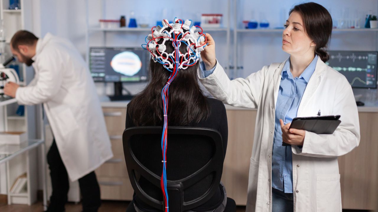
Functional Imaging and Motor Circuit Dysfunction
Functional MRI adds another layer, showing how Parkinson’s disrupts communication between brain regions. Instead of smooth coordination between motor cortex, basal ganglia, and cerebellum, Parkinson’s brains often show inefficient or compensatory activation patterns.
Some regions work overtime to make up for dopamine loss. Others fall out of sync entirely. These network-level disruptions help explain symptoms like bradykinesia (slowness of movement) and freezing of gait.
Functional imaging studies summarized by the National Library of Medicine highlight how Parkinson’s is increasingly understood as a circuit disorder, not just a chemical deficiency (https://www.nlm.nih.gov).
Beyond Movement: Imaging Non-Motor Symptoms
One of the most important contributions of brain imaging research has been validating non-motor symptoms of Parkinson’s. Depression, sleep disturbances, cognitive decline, and autonomic dysfunction aren’t side issues—they’re part of the disease.
Imaging studies show changes in limbic and cortical regions linked to mood and cognition, even in early stages. PET scans reveal altered neurotransmitter systems beyond dopamine, including serotonin and norepinephrine.
This helped dismantle the outdated idea that Parkinson’s is “just” a movement disorder. It’s a multisystem brain disease.
Differentiating Parkinson’s From Look-Alike Disorders
Clinically, Parkinson’s can resemble other conditions like multiple system atrophy (MSA), progressive supranuclear palsy (PSP), or corticobasal degeneration. These disorders respond differently to treatment and progress more aggressively.
Brain imaging helps researchers—and sometimes clinicians—tell them apart. Structural MRI may show characteristic patterns of atrophy in atypical Parkinsonian syndromes. PET and SPECT imaging reveal distinct metabolic and dopamine signatures.
The National Institute on Aging notes that accurate differentiation is crucial for both prognosis and research trials (https://www.nia.nih.gov).
Imaging in Treatment and Drug Development
Modern Parkinson’s drug trials rely heavily on imaging. Dopamine imaging ensures participants actually have dopaminergic degeneration before enrollment. PET scans help measure whether experimental drugs protect neurons or simply mask symptoms.
Imaging is also central to evaluating advanced therapies. Deep brain stimulation (DBS), for example, uses MRI for surgical targeting and functional imaging to study how stimulation reshapes brain networks.
Without imaging, many of these therapies would remain trial-and-error.
Watching the Brain Adapt: Plasticity in Parkinson’s
Despite ongoing neuron loss, the Parkinson’s brain adapts. Imaging studies show compensatory changes, especially early in the disease. Alternative pathways activate to preserve movement and function.
Understanding these adaptive mechanisms has practical consequences. It informs the timing of treatment and rehabilitation, helping clinicians support helpful plasticity rather than disrupt it.
Artificial Intelligence and the Next Wave
AI is increasingly paired with Parkinson’s imaging research. Machine learning models analyze subtle patterns across MRI, PET, and SPECT scans to identify early disease signatures and predict progression.
According to recent NIH-backed reviews, AI-assisted imaging may eventually identify Parkinson’s risk before motor symptoms appear (https://www.nlm.nih.gov). That possibility raises both excitement and ethical questions—especially in a disease without a cure.
What Imaging Still Can’t Do
Despite decades of progress, brain imaging cannot definitively diagnose Parkinson’s on its own. Clinical evaluation remains essential. Imaging findings overlap across disorders and individuals.
Scans also can’t predict exactly how fast someone’s disease will progress or which symptoms will dominate. Parkinson’s remains deeply personal in its course.
The Parkinson’s Foundation emphasizes that imaging supports—but does not replace—clinical judgment (https://www.parkinson.org).
Why Brain Imaging Changed Parkinson’s Research
Brain imaging didn’t eliminate uncertainty in Parkinson’s disease. It clarified it.
It showed that Parkinson’s begins long before tremors, spreads beyond movement circuits, and varies widely from person to person. It shifted research toward early detection, personalized treatment, and realistic expectations.
In Parkinson’s research, imaging isn’t about seeing damage alone. It’s about understanding resilience, compensation, and the slow battle between degeneration and adaptation playing out inside the brain.
That perspective has changed everything.
FAQs:
Can brain imaging diagnose Parkinson’s disease?
No. Imaging supports diagnosis but must be combined with clinical evaluation.
What scan is most specific for Parkinson’s research?
Dopamine transporter PET or SPECT imaging is most informative.
Why is MRI often normal in early Parkinson’s?
Because neuron loss occurs at a microscopic level not visible on standard MRI.